Gallbladder Disease and Gallbladder Endometriosis
Where is the Gallbladder and What does it Do?
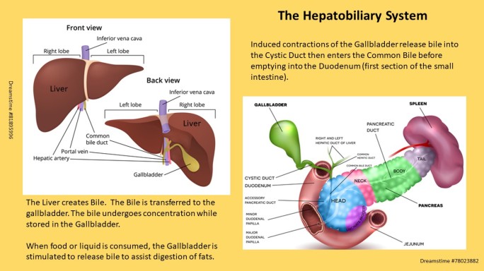
The Gallbladder is an organ of the hepatobiliary system. The liver, bile ducts and gallbladder compose the entire hepatobiliary system. The Gallbladder stores bile produced in the Liver. The bile is condensed after arriving in the gallbladder. Bile aids breaking down consumed fats to be absorbed for use by the body. Bile is about 95% water and 5% dissolved compounds. The number of compounds are extensive but a few important ones include: salts, amino acids, vitamins, bilirubin, phospholipids and cholesterol. The last two compounds (bilirubin and cholesterol) are sensitive to female sex hormones. This hormone sensitivity plays a role in development of gallbladder disease among persons (afab).
In addition to its role in breakdown of ingested fats, the excretion of bile also eliminates toxins, metals and drugs from our body. (1) When a meal is consumed, the gallbladder releases bile into the small intestine (via the Cystic Duct through the Common Bile Duct and into the first section of the Small Intestine (Duodenum) via the Sphincter of Odi. (2) The majority of digestion occurs in this first portion of the small intestine.
Anatomy of the Hepatobiliary System:

Conditions that cause Right Upper Abdominal
(R UQ) Pain
A list (not comprehensive) of some causes for pain in the right upper quadrant:
-
-
-
-
- Liver Diseases
- Gallbladder Diseases
- Colon Cancer
- Stomach and Small Intestine Ulcers
-
-
-
Sex Hormones and Gallbladder disease
The female sex hormones estrogen and progesterone influence the composition of bile and function of the gallbladder. Estrogen can raise the amount of cholesterol in the bile. Elevated cholesterol levels increase bile viscosity (thicker). Elevated levels of progesterone can reduce the forcefulness of gallbladder contractions. Both scenarios impact ability to excrete bile from the gallbladder into the ducts. Hence, both sex hormones, through different mechanisms, increase the risk for gallstone formation.(2)
People (afab) are predisposed to gallbladder disease compared to people (amab) by way of sex hormones. In addition, specific populations exist with even greater risk for gallbladder disease. These include: those who are pregnant (3), use oral contraceptives (risk level dependent upon composition) or take oral hormone replacement.(4)
A significant proportion of women experience biliary disease during pregnancy. Pregnancy may accentuate gallbladder stone formation. Alterations in hepatobiliary function occur during pregnancy to create a lithogenic (stone formation) environment. (3) – Ilhan M et al (2016)
When HRT is applied through transdermal patches, the risk is significantly lower.(5)
Hormone replacement therapy increases the risk of gallbladder disease in postmenopausal women. Use of transdermal estrogens are associated with substantially lower risk of gallbladder disease than use of oral estrogens. (5) – Liu, B et al. (2008)
Common Conditions of the GB and GB Endo Differential Diagnosis
Cholestasis: The reduction or stoppage of bile flow. The skin and whites of the eyes may be discolored yellow, wide spread itching skin, dark urine and light colored stools.(6)
Cholecystitis: Inflammation of the gallbladder. Often occurs when a gallstone blocks the cystic duct for bile to leave the gallbladder. Symptoms often include abdominal pain, nausea or vomiting and, when acute, a fever and/or elevated bilirubin blood values may occur.(6)
Symptoms of gallbladder disease often occur with prolonged periods of fasting or shortly after eating a meal. Meals with higher fat contents aggravate symptoms. (6)
Gallbladder Disease vs Gallbladder Endometriosis
Behaviour of Symptoms:
Gallbladder disease is exacerbated with prolonged fasting or shortly after ingestion of foods and liquids (especially high fat contents). It’s common for resolution of symptoms to occur between episodes and are not associated with the menses.
Both cases of GB Endometriosis (17 yo and 55 yo) reported their episodes to be cyclical and correspond to their menses (catamenial). The 55 year old with GB endometriosis also reported that a constant dull ache present throughout the month, but peaked at menses.
(It is important to note that disease or dysfunctions of numerous structures can create pain in the right upper abdominal quadrant. These structures may also refer pain to the right shoulder. Although both case reports of gallbladder endometriosis did not include these symptoms, the following organs and tissues are among those with this referral pattern:
-
-
-
-
-
-
- Liver
- Gallbladder
- Diaphragm (right)
- Lung (right)
-
-
-
-
-
Blood Draw:
-
- Elevated WBC (inconsistent) = Acute Cholecystitis. Absent with Chronic Cholecystitis.(2)
- All blood values were normal in the two persons with gallbladder endometriosis, with exception the 17 yo adolescent female was anemic.(7,8)
Metabolic Panel:
-
- Elevated Bilirubin = Blocked Bile Duct(s) (often associated with gallstones). (2)
Imaging:
-
- HIDA Scan: Imaging test to examine gallbladder function to contract and expel bile into the ducts. Pathology of the gallbladder (Chronic Cholecystitis) is indicated by an Ejection Fraction (EF) <35%. Conclusive treatment is surgical removal of the gallbladder (cholecystectomy). (2)
- Transcutaneous Ultrasound:
- CT Scan
- MRI
Imaging workup for both cases included Ultrasound and MRI.(7,8) Use of CT with contrast was included in work up for 55 year old due to suspicion of Gallbladder carcinoma with metastasis to abdominal wall.(8)
Although location of symptoms (RUQ), and association with menses (catamenial) were similar among the 17 yo adolescent (7) and 55 yo premenopause people (afab) (8), their diagnostic work up and list of potential causes for their pain differed.
The care team for the 17 year old adolescent person (afab) established a working diagnosis of endometriosis. This occurred, only after exhaustive testing ruled out other causes. The diagnosis of gallbladder endometriosis was established based upon a history of anemia (heavy bleeding with menses), specific appearance during menses and resolution between cycles and confirmed with sequential ultrasound imaging.(7)
Several diagnostic means were used to identify the cause of symptoms, including oesophago-gastroduodenoscopy and colonoscopy as well as radiologic studies. Endscopic examinations showed physiologic findings in the upper and lower gastrointestinal tract. Radiographic examinations, including barium follow-through, initial ultrasound and magnetic resonance imaging (MRI) of the abdomen were normal. Computed Tomography (CT) of the abdomen showed a questionable radiopaque tissue in the wall of the gallbladder, with concomitant inflammation. These findings, combined with the history of complaints, led to the presumed diagnosis of gallbladder endometriosis. US examination was performed once more on d 14 of the menstrual cycle and repeated every second day, showing continual expansion of the suspected tissue. After 12 d and maximum extension of the tissue, a second MRI of the liver was performed, and the diagnosis of endometriosis of the gallbladder could be confirmed. (7)
– Saadat-Gilani K et al (2007)
Although the 55 yo person (afab) also presented with catamenial right upper quadrant pain, presence of two abdominal masses in the area, her age, and presence of chronic, low grade symptoms between peak episodes; resulted in an alternate list of potential diagnoses. These included:
Differential Diagnoses
-
-
-
-
-
- Gallbladder Carcinoma
- Colon Cancer (with metastasis)
- Gallstones
- Chronic Cholecystitis
- Duodenal Ulcer
- Abscess (note: Gallbladder endometriosis – is not on the list)
-
-
-
-
An ultrasound was initially performed of the gallbladder and abdominal wall. A heterogenous mass was reported in the gallbladder and two solid masses within the internal oblique muscle of the abdominal wall. Unable to distinguish these masses as benign or malignant, a CT with contrast was completed. These results were also inconclusive. The final testing, an MRI with a contrast agent used to highlight presence of blood and detailed soft tissue structure was applied to the abdominal masses and gallbladder. The MRI concluded that all masses were low probability as malignant. The definitive treatment was surgical excision of the gallbladder (cholecystectomy) and abdominal masses. The single mass in the gallbladder and two abdominal masses were all endometriosis lesions.(8)
Summary:
Based upon the number of published case studies (2 of 3 known), gallbladder disease has been recorded less than a handful of times. Here we reviewed two of three cases. Both cases presented with pain in the Right Upper Quadrant of the abdomen:
1.) concurrent with the menses. 2.) Absence of symptoms escalation after meals or prolonged fasting.
These are a distinguishing feature between inflammation of the gallbladder and/or presence of gallstones versus endometriosis of the gallbladder.
The age difference between the two cases, and concurrent abdominal masses found in the older person led to alternate list of diagnoses. In regards to the 17 yo.: The negative results of an extensive diagnostic workup and, catamenial nature of the symptoms, a working diagnosis of endometriosis was reached. This case provided ‘text book’ expected findings throughout serial ultrasound imaging from mid month to menses. Sequential imaging over two week period demonstrated progressive enlargement of mass, concurrent with provocation of symptoms. Definitive treatment involved surgical removal of the gallbladder (cholecystectomy).(8)
Citations List: Gallbladder Disease and Endometriosis
To return to Overview GI/Digestive Endometriosis
Looking for a support and education group?
Extrapelvic Not Rare Endometriosis Education and Discussion Group


All Rights Reserved © 2019 Wendy Bingham, DPT. Extrapelvic Not Rare
(last updated 03/06/2021)
