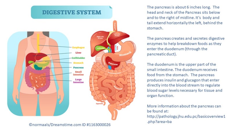
Where is the pancreas and
what does it do?

To date, thirteen cases of pancreatic endometriosis have been published. The first case was presented in 1984. (1) The limited number of publications suggest endometriosis of the pancreas has scant probability of occurrence among persons assigned female sex at birth (afab). However, the ability for it to occur necessitates the disease be recognized and considered when appropriate among persons (afab) with acute pancreatitis or recurrent pain in similar distribution.
Active pancreatic endometriosis has been reported in persons( afab) between 21 and 68 years of age. These included menstruators and post menopause persons (afab), with and without a history of endometriosis. (1-10) In a single case, repetitive, cyclical symptoms was associated with menses (catamenial). (5) Others reported symptoms concurrent with acute pancreatitis or in the months following. (1,2,7,8). A few cases denied an episode of acute pancreatitis but reported low level of recurring symptoms over the previous year. (3,5,6)

Symptoms
The following symptoms were noted among the 13 reported cases:
- (n=13) Epigastric pain (1-10)
(1) Specified left side epigastric pain (3)
*(1) Radiation into the Left chest and back (5)
- (2) Weight Loss (#15/prior 3 months and #20/prior year) (2,6)
- (1) Nausea and Vomiting (8).
- (1) Left Loin Pain (the pancreatic lesion surrounded the pancreatic tail and kidney) (11)
- (1) Diarrhea (2)
- (1) Palpable mass in left upper abdomen (7)
- (1) Cyclic, recurrent symptoms 7 days prior to menses (catamenial) (5)
- (1) No relationship to menses or symptoms of endometriosis elsewhere (4)
Blood Draw
Blood is drawn to analyze values which help differentiate various disease processes. No patterns of were observed among those with endometriosis lesions of the pancreas. Results of blood tests varied. Among reported findings include:
Amylase High (2) Normal (5)
Carcino-Embryonic Antigen High (7) Normal (2,3) (CEA-Cancer Antigen 19-9)
CA-125 High (7) Normal (3)
Tumor Markers Negative (5)
Lipase Normal (5)
C-Reaction Protein Normal (5)
GGT, ALAT and CRP High (3)
Imaging
Imaging is a useful tools to assist in diagnostics. However, like many areas of the body with endometriosis, imaging is not sensitive enough to detect the presence of all endometriosis. It also has limitations to identify all endometriosis lesions from other lesions. These limitations also apply to imaging of the pancreas. It can be challenging to determine whether a cyst is benign, pre-malignant or malignant. Among the 13 cases, a single study made was able to make a ‘preliminary diagnosis’ of endometriosis based upon imaging and lab values pre-operatively. (4) The remaining twelve cases could not definitively conclude that the lesion(s) under assessment were not premalignant or malignant from imaging scans . In these 12 cases, all underwent surgery to remove part of the pancreas, and other tissues as appropriate.
The single case study which reached a preoperative diagnosis involved tissue surrounding the pancreas (not within the body or tail of the pancreas) and were able to perform serial imaging with both CT and MRI. (4). Based upon analysis across time, alterations in lesion size and findings on MRI supported evidence of hemorrhagic fluids which suggested endometriosis and not one of the two malignant neoplasm: Psuedopapillary tumor and Mucinous Cystic Neoplasm (MCN). In this case, the surgical team was able to remove the lesion without resection of the pancreas, spleen or other structures.
‘Traditional’ characteristics of endometrioma on imaging scans:
The ‘traditional’ image of an endometrioma is:
- unilocular (single room)
- homogeneous without septations (analogy: house without interior walls that divide into rooms).
Less frequent, they may present with:
- multiple loculi (analogy: grapes on a vine)
- septations (A house with internal walls; more walls equal more of rooms)
- contain solid components and nodules. (11)
‘Non-traditional’ Imaging characteristics among pancreatic endometriosis cases:
The variation of characteristics is observed among the Computer Tomography (CT) and Ultrasound (US) images of those with pancreatic endometriosis:
Computer Tomography (CT)
loculated and thickened’ (2)
cystic, solid component with calcification; with contrast: partial cystic and partial hemorrhagic (3)
capsular, possible septations (6)
Ultrasound (US)
septate walls and mural nodules (5)
Following their case analysis and literature review of previous publications, the authors of a 2016 case summarized the following constraints of imaging and ability to distinguish endometriosis from other types of cysts:
“Since there are only few reports regarding endometrial cysts (in the liver) or pancreas, no typical imaging features have been established. A common feature is a cystic lesion, sometimes with hemorrhagic content and in a few cases also with a solid component. In our case, the CT Scan showed a cystic lesion with a seemingly solid portion and a small calcification. The MRI scan demonstrated a partly serous, partly hemorrhagic lesion in the pancreatic tail with only minimal contrast enhancement of the cyst wall. The hemorrhage component might have raised the suspicion of endometriosis, but the finding remains unspecific. Since these imaging findings are nonspecific, a definite diagnosis is difficult to establish and a comprehensive work up regarding the possible malignant features, tumor markers, fine needle aspiration/biopsy, a previous history endometriosis as well as a pancreatitis and possible menstrual cycle dependency of symptoms or changing imaging features over time can be helpful in establishing a diagnosis” – Plodeck V et al. (2016) (3)
More details about imaging characteristics with Ultrasound and Magnetic Resonance Imaging (MRI) are located at https://radiopaedia.org/articles/endometrioma. (11) (Note: this link is to be used for educational purposes only. It is not intended for self-diagnosis).
Treatment
If blood tests, imaging, guided-needle biopsy (as appropriate), and absence of cyclic catamenial recurrence, are not able to provide a clear distinction between a benign and premalignant or malignant cyst, resection of the involved pancreatic tissue (and possibly adjacent structures like the spleen is recommended to ensure lesion is not malignant. Twelve of the thirteen published cases underwent resections.
Endometriosis cysts have been identified in body and tail of the pancreas.

A variety of benign and malignant cysts can form within the pancreas. A very common cyst which appears about 4 to 8 weeks after an initial Acute Pancreatitis is a Pseudocyst. Pseudocysts are most common around the Head of the Pancreas and form as a result of lingering inflammation. (12) These are benign. It is important to note these cysts are not present in imaging during an initial Acute Pancreatitis.
One specific type of cyst which is found among persons (afab), more common middle-age upward are Mucinous Cystic Neoplasms (MCN). These neoplasms have characteristics similar to the ovaries. Like endometriosis, MCN cysts are often responsive to Estrogen and/or Progesterone hormones. Unlike endometriosis, Mucinous Cystic Neoplasms (MCN) are (+) for Inhibin. (13)
“The findings of Alpha-Inhibin positivity in MCN with ovarian-type stroma further supports its similarity to true ovarian stroma tissue and may suggest a role of complex hormonal interaction in the pathogenesis”. (11) – Yen MM et al. (2004)
Summary
Since 1984, thirteen cases of pancreatic endometriosis has been published. These lesions have been found in persons (afab) with and without a history of endometriosis; pre and post menopause age; with and without history of Acute Pancreatitis. To date there have been no consistent blood values, markers or image findings that can definitively determine endometriosis versus a premalignant or malignant cyst. (The case study which considered endometriosis as a preoperative diagnosis involved a peri pancreatic lesion that did not occupy the parenchyma of the pancreas. They also had opportunity to use serial imaging over a period of time to monitor changes in the lesion based upon significantly reduced symptomology after the initial acute presentation and low probability lesion was malignant that allowed a five month delay from acute presentation to surgical intervention). All cases reported epigastric pain, no lesions were found ‘incidentally’. The ability to differentiate endometriosis from other cystic lesions of the pancreas is complicated further by its heterogeneous appearance on imaging.
Endometriosis of the Pancreas Citations
To return to Overview GI/Digestive Endometriosis
Extrapelvic Not Rare Endometriosis Education and Discussion Group


All Rights Reserved © 2019 Wendy Bingham, DPT. Extrapelvic Not Rare
