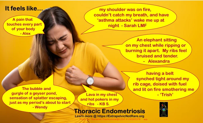
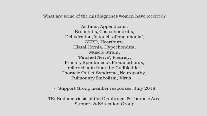
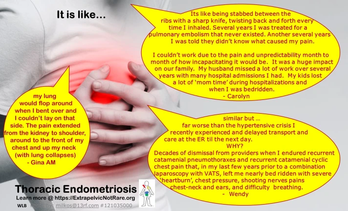
1.) Thoracic Endometriosis Syndrome (TES) involves any tissue or organ in the chest cavity and diaphragm. (1,2,3)
2.) Most to least common location of lesions found in the respiratory system (1,2,3,4,)
- Diaphragm (abdominal>chest side)
- Parietal Pleura (Chest Wall and surface of diaphragm,chest side)
- Visceral Pleura (surface covering the lungs)
- Lung Parenchyma (lung & air exchange areas) Bronchi.
3.) No single theory of pathogenesis explains all locations and presentations of endometriosis found in the respiratory system (3).
4.) Recurrent cyclical chest pain is the most common initial presentation of TE. A smaller portion of sufferers experience these manifestations, in descending frequency (1,2):
- Catamenial Pneumothorax (more below)
- Catamenial Hemothorax
- Catamenial Hemoptysis (more below)
- Lung Nodules
(Clarification: A Manifestation can be measured and recorded through imaging, blood tests etc to verify an abnormal condition is present (a type of ‘sign’ – can be ‘visualized’). A symptom is a persons perception of an event that cannot be measured with imaging, blood tests etc but can give a subjective scale of its intensity, location and duration (ie. sensations of burning, weakness, achy, feel shortness of breath or difficulty breathing).
5.) The respiratory system is the third most frequent body system affected by extra-pelvic endometriosis lesions. The digestive and urinary/excretory systems are the two more commonly affected than respiratory (5).
6.) Symptoms most often begin to appear around menses. As disease progresses, it may include mid-month (ovulation), expanding to 24/7 symptoms with peaks at menses and ovulation.
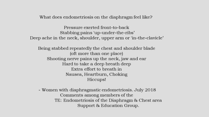
Are you, or someone you know experiencing symptoms that occur around your menses that give’s suspicion for thoracic endometriosis?
Please click on the link below to learn more about the disease, what to monitor and how to advocate for yourself when seeking care from a provider:
TE Patient Perspectives and Tools
The fact TE is under recognized, under diagnosed and the ability to document all persons with the disease is a yet to occur, it is unclear the true probability that those who present with cyclic, catamenial chest pains develop manifestations. Just because a person has not developed a pneumothorax, pleural effusion or coughed blood – does NOT exclude TE from being present. A person CAN be symptomatic WITHOUT manifestations.
Patient Perspective:
I saw a new doctor today and I mentioned that I had diaphragmatic endometriosis. “That’s incredibly rare,” he said. “That’s an impressive doctor you have that made that diagnosis.” But here’s the thing…My doctor didn’t make that diagnosis, I did. I had problems breathing for years. I had to yawn to take a deep breath & would have sharp shooting chest pains that would literally leave me breathless.
I saw pulmonologists & one even gave me an inhaler. But nothing appeared wrong. So I did some research and thought, maybe it’s endometriosis on my diaphragm. I already had 4 surgeries for fairly widespread endo, so didn’t seem far off. I told the gyn doing my next surgery my theory and she laughed in my face, “That’s really rare, it’s likely just stress.”
So I paid an extra $1,000 to have the specialist who trained her supervise my surgery, and I begged them to look at my diaphragm. “Please, just while you’re in there.” When I woke up from surgery it was the first time in my memory that I could take a deep breath. I cried.
I had the largest piece of diaphragmatic endometriosis my doctor had seen. “It’s a good thing we looked,” she said. But none of that would’ve happened if I hadn’t spent more than a year insisting. So yeah, maybe it’s impressive that my doctor saw & removed it. But I diagnosed it. – Jordan (2018)
Symptoms of disease in the diaphragm and chest cavity can progress for many years before a manifestation occurs. Manifestations are dependent upon: lesion location and aggression of lesions. Is it appropriate for a person to potentially suffer for many years with symptoms before there is willingness among providers to ‘look’ for disease if all other possible causes have been ruled out?
The various mechanisms of CP are discussed below in ‘How does endometriosis create a Catamenial Pneumothorax?’.
As the most frequent manifestation of TES, (about 3/4 of those who develop pathology that can be identified on imaging are Seconday Spontaneous Pneumothoraxes referred to as Catamenial Pneumothorax CP or Thoracic Endometriosis Related Pneumothorax TERP) its important do understand what it feels like to develop a Catamenial Pneumothorax. Not ever person’s experience is the same.
A member of our TE community who experienced recurrent CP’s gathered data from members of our support group community. To view the variety of symptoms, clinical findings and interventions please click on the following link: CatamenialPneumothorax-SurveyofWomenwhoexperienced1ormoreLungCollapsesconfirmedbyMedicalImaging
It is vital to identify the potential of endometriosis as a cause for recurrent spontaneous pneumothoraxes. If investigation of the patients past medical history, and investigation of diagnosed or symptoms of undiagnosed pelvic endometriosis, history of cyclic scapula/chest pains concurrent with menses, misdiagnosis of Primary SPT can lead to interventions to prevent recurrence with disease unidentified and left behind. Please click on the link here to view a case example:
How does endometriosis create a Catamenial Pneumothorax?
There are multiple ways endometriosis can cause a lung collapse. Lesion location(s) determine the way in which a lung collapses. (1)
Diaphragm Lesions:
a.) Lesions found on the diaphragm may progress to ‘full thickness’ that leads to holes or fenestrations through the diaphragm. This allows air to pass between the abdominal cavity and the chest cavity. Under normal circumstances there is minimal air between the chest wall and the lung (in fact a negative pressure value normally occurs to help keep the lung expanded). At time of menses, a woman’s cervix opens to allow the debri of menses to be expelled from the body. It is postulated that air freely enters through the cervix and up into the abdomen through the open ended Fallopian tubes. This results in positive air pressure which passes into the chest cavity. The pressure difference results in air pressing inward on the lung that results in collapse of lung inward.
Visceral Pleura (outer surface of the lung):
b.) Lesions on the outer lung surface (visceral pleura) create weakness in the lung wall. Air exchange occurs in peripheral lung tissue up to the lung wall. As a lesion deepens, it penetrates into the lung tissue. Because the pressure outside of the lung, but inside the chest cavity is lower than pressure inside the lung, air moves from inside the lung to the chest cavity.escapes into the lower pressured air in the chest cavity.
Parenchyma/Bronchiole Lesions:
c.) Lesions located in the lung parenchyma (inside the lung) may result in various presentations. As noted above, lesions in larger airways or in lung tissue close to an airway can cause a person to cough blood at menses (catamenial hemoptysis). Lesions found among the air exchange regions may present with shortness of breath and/or difficulty breathing (dyspnea). Lesions located in the peripheral regions of the lung can lead to blebs or bulla formation if the lesions cause structural changes. If endometriosis cells are located in the passageways where air enters into air exchange areas, they may swell and create a ‘check-valve’ mechanism in which air enters into the lung tissue but the lesions restrict escape of air (that occurs with exhalation). This leads to overinflation of air exchange regions. As a result, Blebs and Bulla form and may rupture – inducing a pneumothorax (1). Poh et al (2) describe Lillington’s 1972 theory as follows:
The swelling of intrapulmonary endometrial implants along with premenstrual hormonal changes cause an obstruction of terminal bronchioles with localised hyperinflation and rupture of distal alveoli. – Poh CL et al (2011)
d.) Lesions of the chest wall (parietal pleura) may create bloody pleural effusion (hemothorax) or, if adhered to the outer surface of the lung wall (visceral pleura) may penetrate or avulse to induce a pneumothorax similar to (b) above.
Citation: TEReferencesHowEndoCreatesCP
Are there ‘clues’ to suggest who may have TE?
My long journey to find answers and a care team who did not dismiss me, imply I was exaggerating or refused to see the connection between the symptoms I presented with, which were ‘atypical’ but ‘catamenial’, caused me to put pen-to-paper for others who struggle to be heard or are unsure if they have the disease.
The articles I have written provide an overview about thoracic endometriosis, other diseases that must be considered and tools to help a person who may be suffering from this disease, for better advocacy and documentation of the experience. Two important factors: Self-Advocacy and locate an experienced multidisciplinary surgical team with practical experience treating this specific region. To access this article ‘TE Patient Perspective and Tools’ please click here:
TE Patient Perspectives and Tools
In 2011, Roussett-Jablonski et al created a Table of Clinical Patterns for Diagnosis of Thoracic Endometriosis. Measurements of medical history and specific symptoms were aligned with the presence or absence of confirmed endometriosis. The Sensitivity and Specificity of each item measured and likelihood TE occurred was created. This chart was based upon the results of 156 subjects over 8 yrs. To view the full article (see Table 1), please click here: Roussett-Jablonskietal.2011Table1
The manifestations are proof of disease, but histological confirmation can be difficult to obtain. – Flavio Colaut et al. (2017)
Despite the fact that disease can cause lungs to collapse, the chest cavity to fill with bloody-pleural effusions or expectorate sputum with various amounts of blood, finding ‘concrete’ proof of the cause is not easy. Variability to acquire histological confirmation of endometriosis falls widely on the spectrum between (est 30-70% operative cases) (1-7) but other associated evidence of active disease is observed intraoperatively. Signs of prior insults include blebs, adhesions, scarring, discoloration etc.
Flavio Colaut et al. describe the diagnosis of thoracic endometriosis based upon the ‘Ex Juvantibus Principle’ . A diagnosis is reached through clinical reasoning. The concurrence of menses at the time of event, with a repetitive, cyclic occurrence lends to a differential diagnosis of ‘catamenial’. The use of medical management which may disrupt the cyclical characteristic supports the diagnosis (8).

Citations List: TEReferencesAreThereClues
Catamenial Hemoptysis
A small portion of those with Thoracic Endometriosis cough varying degrees of blood-tinged sputum at the time of menses. A compilation of case studies, their treatments and self-help suggestions to prepare for discussion with your healthcare team can be accessed here: CoughingUpBloodWhoCanHelpandCaseStudies
Support and Education Group for Sufferers and Healthcare Providers
If you are a person diagnosed with/or concerned you may have disease of the respiratory system, support someone with the disease or a healthcare provider who wants to learn more about the condition from those with the disease and/or specialists who surgically treat disease of the thoracic cavity, please join us at:
Extrapelvic Not Rare Endometriosis Education and Discussion Group

Tweet to @XtraNotRarehttps://platform.twitter.com/widgets.js
All Rights Reserved. © 2018 – Wendy Bingham, DPT

